Examinations

Optical Examination

Two complementary optical methods — Wood's Lamp and dermoscopy — support pigment assessment, lesion evaluation, and treatment monitoring.

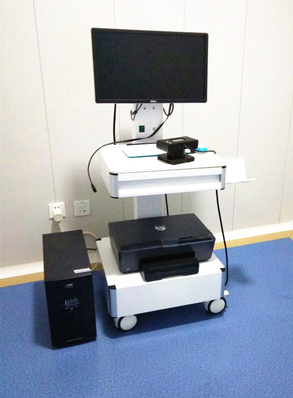
Wood's Lamp examination workstation

Wood's Lamp

Ultraviolet Fluorescence Examination

Wood's Lamp emits long-wave ultraviolet (UVA) light, allowing differences in skin pigmentation and fluorescence to become more visible.

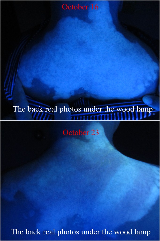
Under Wood's Lamp examination, areas of reduced or absent pigmentation can appear more clearly defined, helping clinicians assess the extent and distribution of depigmentation.

Key Applications

  • Identify subtle depigmented areas
  • Define lesion borders
  • Assess residual pigmentation
  • Compare pigmentation before and after treatment
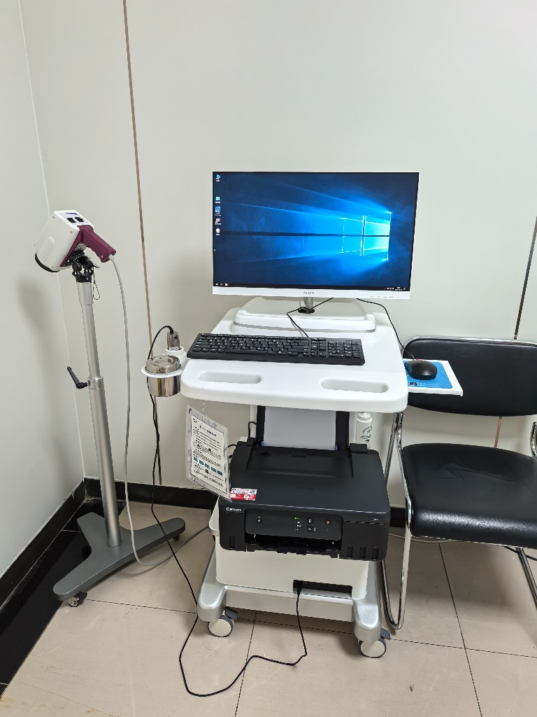
Dermoscopy examination workstation

Dermoscopy

Magnified Skin Structure Analysis

Dermoscopy provides a magnified view of skin structures that may not be clearly visible to the naked eye.

It allows clinicians to examine pigment patterns, perifollicular changes and other surface or subsurface features, providing additional information for clinical assessment.

Key Applications

  • Examine pigmentation patterns
  • Assess perifollicular pigmentation
  • Observe subtle skin changes
  • Support diagnosis and treatment monitoring

Principle Comparison

Wood's Lamp

UV Light → Skin Fluorescence → Pigment Contrast

Wood's Lamp enhances the visual contrast between normally pigmented and depigmented skin, making subtle areas of pigment loss easier to identify.

Dermoscopy

Magnification → Structural Visualization → Detailed Assessment

Dermoscopy magnifies the skin surface and reveals pigmentary and vascular structures that may not be visible during routine examination.

Treatment Progress Under Wood's Lamp

Visualizing Changes in Skin Repigmentation

Wood's Lamp before-and-after comparison of treatment progress